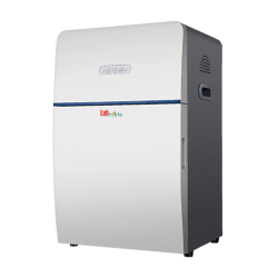
Chemiluminescence Imaging System LMCIS-A101 features an exceptionally high quantum efficiency CCD Camera with a sensor of size 12.49 × 9.99 mm, providing enhanced sensitivity and an extended dynamic range for imaging chemiluminescence gels. Offers high-resolution imaging capabilities with its F0.95 motorized lens, allowing for detailed visualization of chemiluminescent signals. Incorporates automated exposure control for precise and consistent imaging across different samples.
Get Quote| CCD Sensor Size | 12.49 × 9.99 mm |
| Resolution | 6.05 Megapixels, 2750 × 2200 |
| Pixel Density | 16bit (65536 Grey Scales) |
| Pixel Size | 4.54 × 4.54 µm |
| Lens | F0.95 motorized lens |
| Light Source |
LED Epi-white light × 2 UV-Transilluminator (302 nm) White-LED Transilluminator |
| Display | NA |
| Quantum Efficiency | ≥ 75 % |
| Readout Noise | < 5.5e- RMS |
| Dark Current | 0.0003 e/p/s |
| Dynamic Range | 4 orders of magnitude |
| Standard Filter | 590 nm filter |
| Filter Wheel | 8 Sockets Filter Wheel |
| Max Image Area | 260 × 210 mm |
| Cooling Temperature | -30˚C |
| Software | Image acquisition and analysis software |
| Internal Dimension | 380 × 350 × 490 mm |
| External Dimension | 560 × 480 × 780 mm |
| Weight | 39 kg |
 CCD camera exhibits a remarkable ≥ 75% high quantum efficiency
 Provides user-friendly image navigation and browsing for convenient operation
 Allows optional attachment of different fluorescent light sources and filters
 Utilizes automatic pixel binning technology for enhanced imaging efficiency
 Enables marker image capture and composition with objective bands
 Occupies a small footprint, minimizing bench space requirements
 Maximum sensitivity configuration
 Optional System Configuration of RNA, DNA, Protein, Chemiluminescence, Fluorescence System
 High-Precision Automatic Exposure
 One-Touch Image Acquisition
 Marker Image Capture and Composition
 Automatic Capture Parameters Access
 Convenient Image Navigation
 Image Rotation, Cropping, and Counter Color Processing
 Advanced Pixel Binning Technology
 Optional Excel File Save for Analysis Results
 Optical Density Calculation
 Background Mode Optimization
It is employed in genetic engineering, biotechnology, and molecular biology to capture high-quality images of DNA/RNA bands. It is also utilized for protein detection, ELISA plate reading, and comprehensive Chemiluminescence analysis.
Get Quote for
Chemiluminescence Imaging System LMCIS-A101