Get Quote
Contact us now to receive a detailed quotation.
Overview
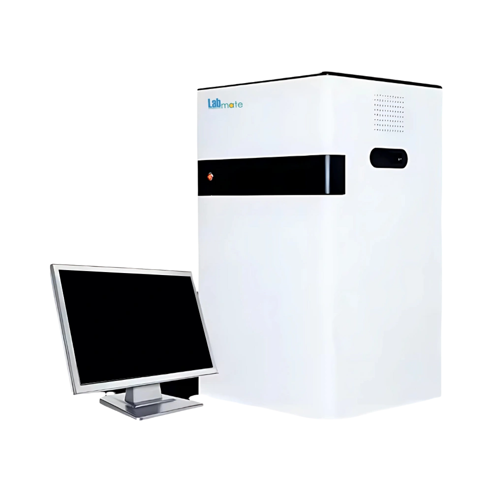
Gel Documentation System LMGDO-A100 captures and documents DNA, RNA, and protein gels using a 5-megapixel camera, motorized F1.2 lens, and 302 nm UV transmission. Binning modes, multi-frame accumulation, automated calculations, and Excel export support gel analysis and reporting. It is suited to molecular biology, genetics, biochemistry, and electrophoresis workflows.
Features :
Specifications :
| Camera Type | Digital Camera |
| Resolution | 5 Megapixels (2592 × 1944) |
| Dynamic Range | 4.8 OD |
| Pixel Density | 16-bit (65,536 Grey Scales) |
| Lens | Motorized Lens F1.2 |
| UV Transmission | 302 nm |
| UV Transmission area | 21 × 26 cm |
| White LED Transmission area | 19 × 26 cm |
| Dimensions | 630 × 560 × 900 mm |
| Weight | 50 Kg |
Optional Accessories :
Model No | Accessory |
LMGDO-A100-B1 | Blue LED transmission |
Applications :
Gel Documentation System LMGDO-A100 supports imaging and documenting DNA/RNA gels, protein electrophoresis, and chemiluminescence with in academic, research, and clinical laboratories.
Selection Guide :
Gel Documentation System LMGDO-A100 is an essential imaging solution designed for laboratories involved in molecular biology, genetics, and biochemistry research. It is primarily used for visualizing, documenting, and analyzing nucleic acid and protein gels stained with dyes such as ethidium bromide, SYBR Safe, or Coomassie blue. This system is suitable for DNA/RNA electrophoresis analysis, protein gel imaging, and fluorescent or chemiluminescent detection in a variety of experimental settings. The model supports high-resolution imaging and offers reliable documentation of gel results, making it suitable for academic, clinical, and pharmaceutical research. Additionally, the system often complies with CE and ISO certifications, ensuring it meets international standards for quality and safety. Researchers can select this model based on its application compatibility, high sensitivity imaging, and validated performance in peer-reviewed studies and laboratory workflows.